The Effect of Ginkgo Biloba Extract on Mice Treated with Taxol
DOI:
https://doi.org/10.51173/ijmhs.v2i2.62Keywords:
Ginkgo Biloba, Mice , Taxol , Diabetes Mellitus , CancerAbstract
Background: The Ginkgo biloba plant is the sole extant species of the Ginkgoaceae family and is among the oldest seed plants. The extract of Ginkgo biloba leaves can also augment the activities of antioxidant enzymes, including both enzymatic and nonenzymatic systems. Antioxidant enzymes comprise catalase, superoxide.
Objective of study: Thirty-six albino mice, sourced from Babylon University and weighing 30±5 grams, were utilized in this study. Water and processed dry food were provided. The animals were categorized into six groups, with six mice per group, and maintained under standardized circumstances (25°C, 12-hour light/12-hour dark cycle).
Materials and Methods: In this study, 100 pregnant women—50 PCS sufferers and 50 stable controls—were assessed for functional polymorphisms of the TGFB1 gene [C-509 T]. Heterozygous CT accounted for 24% of PCS patients and 30% of controls, whereas homozygous CC accounted for 76% of PCS cases and 70% of normal, healthy pregnant women.
Results: Our phytochemical analysis of methanol-aqueous Ginkgo biloba leaf extracts revealed the presence of essential oils, alkaloids, flavonoid glycosides, tannins, phenolic compounds, saponins, coumarins, and terpenes. In contrast, steroids and resin exhibited poor results.
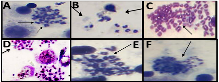
Conclusion: This study showed that Taxol (6 mg/kg b.wt.) generated cytotoxicity and genotoxicity in the liver and spleen; however, these adverse effects may be mitigated or even prevented in certain instances by the protective effect.
References
References
Teng, M. L., Ng, C. H., Huang, D. Q., Chan, K. E., Tan, D. J., Lim, W. H., ... & Muthiah, M. D. (2022). Global incidence and prevalence of nonalcoholic fatty liver disease. Clinical and molecular hepatology, 29(Suppl), S32. https://doi.org/10.3350/cmh.2022.0365.
Riazi, K., Azhari, H., Charette, J. H., Underwood, F. E., King, J. A., Afshar, E. E., ... & Shaheen, A. A. (2022). The prevalence and incidence of NAFLD worldwide: a systematic review and meta-analysis. The lancet gastroenterology & hepatology, 7(9), 851-861. https://doi.org/10.1016/S2468-1253(22)00165-0.
Friedman, S. L., Neuschwander-Tetri, B. A., Rinella, M., & Sanyal, A. J. (2018). Mechanisms of NAFLD development and therapeutic strategies. Nature medicine, 24(7), 908-922. https://doi.org/10.1038/s41591-018-0104-9.
Rinella, M. E., Neuschwander-Tetri, B. A., Siddiqui, M. S., Abdelmalek, M. F., Caldwell, S., Barb, D., ... & Loomba, R. (2023). AASLD Practice Guidance on the clinical assessment and management of nonalcoholic fatty liver disease. Hepatology, 77(5), 1797-1835. https://doi.org/10.1097/HEP.0000000000000323.
Méndez-Sánchez, N., Bugianesi, E., Gish, R. G., Lammert, F., Tilg, H., Nguyen, M. H., ... & Awolowo, O. (2022). Global multi-stakeholder endorsement of the MAFLD definition. The lancet Gastroenterology & hepatology, 7(5), 388-390. https://doi.org/10.1016/S2468-1253(22)00062-0.
Lonardo, A., Bril, F., Caldwell, S. H., Eslam, M., Fan, J. G., Gish, R. G., ... & George, J. (2024). Researchers call for more flexible editorial conduct rather than abruptly adopting only the new MASLD nomenclature. Journal of Hepatology, 80(5), e192-e194. https://doi.org/10.1016/j.jhep.2024.01.012.
Rinella, M. E., Lazarus, J. V., Ratziu, V., Francque, S. M., Sanyal, A. J., Kanwal, F., ... & NAFLD Nomenclature Consensus Group. (2023). A multisociety Delphi consensus statement on new fatty liver disease nomenclature. Hepatology, 78(6), 1966-1986. https://doi.org/10.1097/HEP.0000000000000520.
Younossi, Z. M., Paik, J. M., Stepanova, M., Ong, J., Alqahtani, S., & Henry, L. (2024). Clinical profiles and mortality rates are similar for metabolic dysfunction-associated steatotic liver disease and non-alcoholic fatty liver disease. Journal of Hepatology, 80(5), 694-701. https://doi.org/10.1016/j.jhep.2024.01.014.
Bassal, T., Basheer, M., Boulos, M., & Assy, N. (2022). Nonalcoholic fatty liver disease—a concise review of noninvasive tests and biomarkers. Metabolites, 12(11), 1073. https://doi.org/10.3390/metabo12111073.
Ramírez-Mejía, M. M., Jiménez-Gutiérrez, C., Eslam, M., George, J., & Méndez-Sánchez, N. (2024). Breaking new ground: MASLD vs. MAFLD—which holds the key for risk stratification? Hepatology international, 18(1), 168-178. https://doi.org/10.1007/s12072-023-10620-y.
Kouvari, M., Valenzuela-Vallejo, L., Guatibonza-Garcia, V., Polyzos, S. A., Deng, Y., Kokkorakis, M., ... & Mantzoros, C. S. (2023). Liver biopsy-based validation, confirmation and comparison of the diagnostic performance of established and novel non-invasive steatotic liver disease indexes: Results from a large multi-center study. Metabolism, 147, 155666. https://doi.org/10.1016/j.metabol.2023.155666.
Campos-Murguía, A., Ruiz-Margáin, A., González-Regueiro, J. A., & Macías-Rodríguez, R. U. (2020). Clinical assessment and management of liver fibrosis in non-alcoholic fatty liver disease. World journal of gastroenterology, 26(39), 5919. https://doi.org/10.3748/wjg.v26.i39.5919.
Cataldo, I., Sarcognato, S., Sacchi, D., Cacciatore, M., Baciorri, F., Mangia, A., ... & Guido, M. (2021). Pathology of non-alcoholic fatty liver disease. Pathologica, 113(3), 194. https://doi.org/10.32074/1591-951X-242.
Yu, J. H., Lee, H. A., & Kim, S. U. (2022). Noninvasive imaging biomarkers for liver fibrosis in nonalcoholic fatty liver disease: current and future. Clinical and molecular hepatology, 29(Suppl), S136. https://doi.org/10.3350/cmh.2022.0436.
Nogami, A., Yoneda, M., Iwaki, M., Kobayashi, T., Honda, Y., Ogawa, Y., ... & Nakajima, A. (2022). Non-invasive imaging biomarkers for liver steatosis in non-alcoholic fatty liver disease: present and future. Clinical and molecular hepatology, 29(Suppl), S123. https://doi.org/10.3350/cmh.2022.0357.
Jang W, Song JS. Non-invasive imaging methods to evaluate non-alcoholic fatty liver disease with fat quantification: a review. Diagnostics. 2023;13(11):1852. https://doi.org/10.3350/cmh.2022.0357.
Jang W, Song JS. Non-invasive imaging methods to evaluate non-alcoholic fatty liver disease with fat quantification: a review. Diagnostics. 2023;13(11):1852. https://doi.org/10.3390/diagnostics13111852.
Trujillo MJ, Chen J, Rubin JM, Gao J. Non-invasive imaging biomarkers to assess nonalcoholic fatty liver disease: A review. Clin Imaging. 2021; 78:22–34. https://doi.org/10.1016/j.clinimag.2021.02.039.
Zhou JH, Cai JJ, She ZG, Li HL. Noninvasive evaluation of nonalcoholic fatty liver disease: Current evidence and practice. World J Gastroenterol. 2019;25(11):1307. https://doi.org/10.3748/WJG.V25.I11.1307.
Rabah, S. O., Ali, S. S., Alsaggaf, S. M., & Ayuob, N. N. (2010). Acute taxol toxicity: the effects on bone marrow mitotic index and the histology of mice hepatocytes. Journal of Applied Animal Research, 38(2), 201-207.https://doi.org/10.1080/09712119.2010.10539511.
Bril, F., McPhaul, M. J., Caulfield, M. P., Castille, J. M., Poynard, T., Soldevila-Pico, C., ... & Cusi, K. (2019). Performance of the SteatoTest, ActiTest, NashTest and FibroTest in a multiethnic cohort of patients with type 2 diabetes mellitus. Journal of Investigative Medicine, 67(2), 303-311. https://doi.org/10.1136/jim-2018-000864.
Chen, L. W., Huang, P. R., Chien, C. H., Lin, C. L., & Chien, R. N. (2020). A community-based study on the application of fatty liver index in screening subjects with nonalcoholic fatty liver disease. Journal of the Formosan Medical Association, 119(1), 173-181. https://doi.org/bham-ezproxy.idm.oclc.org/10.1016/j.jfma.2019.03.016.
Rossi, R., De Palma, A., Basilico, F., & Mauri, P. (2011). Analytical methods for characterizing bioactive terpene lactones in ginkgo biloba extracts and performing pharmacokinetic studies in animal and human. INTECH Open Access Publisher. https://doi.org/10.5772/13068.
Sadowska-Krępa, E., Kłapcińska, B., Pokora, I., Domaszewski, P., Kempa, K., & Podgórski, T. (2017). Effects of six-week Ginkgo biloba supplementation on aerobic performance, blood pro/antioxidant balance, and serum brain-derived neurotrophic factor in physically active men. Nutrients, 9(8), 803.https://doi.org/10.3390/NU9080803.
Serrano-García, N., Pedraza-Chaverri, J., Mares-Sámano, J. J., Orozco-Ibarra, M., Cruz-Salgado, A., Jiménez-Anguiano, A., ... & Trejo-Solís, C. (2013). Antiapoptotic effects of EGb 761. Evidence‐Based Complementary and Alternative Medicine, 2013(1), 495703.https://doi.org/10.1155/2013/495703.
Han, A. L., & Lee, H. K. (2022). Comparison of the diagnostic performance of steatosis indices for discrimination of CT-diagnosed metabolic dysfunction-associated fatty liver disease. Metabolites, 12(7), 664. https://doi.org/10.3390/METABO12070664.
Flessa, C. M., Kyrou, I., Nasiri‐Ansari, N., Kaltsas, G., Kassi, E., & Randeva, H. S. (2022). Endoplasmic reticulum stress in nonalcoholic (metabolic associated) fatty liver disease (NAFLD/MAFLD). Journal of cellular biochemistry, 123(10), 1585-1606. https://doi.org/10.1002/JCB.30247.
Flessa, C. M., Kyrou, I., Nasiri-Ansari, N., Kaltsas, G., Papavassiliou, A. G., Kassi, E., & Randeva, H. S. (2021). Endoplasmic reticulum stress and autophagy in the pathogenesis of non-alcoholic fatty liver disease (NAFLD): current evidence and perspectives. Current obesity reports, 10(2), 134-161. https://doi.org/10.1007/s13679-021-00431-3.
Huang, X., Zhang, X., Song, J., Lan, D., Wang, M., Ji, X., ... & Meng, R. (2025). The mechanisms of efficacy and safety of Ginkgo biloba extract in acute ischemic stroke: a real-world study. Thrombosis Journal, 23(1), 1-10. doi: 10.1186/s12959-025-00696-x.
Al-Ameedi, A. I., Ayad, Z. M., Mohammed, W. A., & Hajwal, S. K. (2023). Ginkgo biloba extract’s efficacy to mitigate the genotoxicity that hydroxyurea induces in mice. Adv. Anim. Vet. Sci, 11(4), 552-557. DOI:10.17582/journal.aavs/2023/11.4.552.557.
Mulianto, N., Kariosentono, H., Purwanto, B., Indarto, D., & Soetrisno, S. (2024). The Effect of Ginkgo Biloba Extract on Oxidative Stress, Anti-Inflammatory and Antiapoptotic in Vitiligo Treated with Topical Desoxymethasone. EVOLUTIONARY STUDIES IN IMAGINATIVE CULTURE, 1580-1590. DOI: https://doi.org/10.70082/esiculture.vi.1483.
Singh, S. K., Srivastav, S., Castellani, R. J., Plascencia-Villa, G., & Perry, G. (2019). Neuroprotective and antioxidant effect of Ginkgo biloba extract against AD and other neurological disorders. Neurotherapeutics, 16(3), 666-674. https://doi.org/10.1007/s13311-019-00767-8.
Abdel-Fatah, S. S., El-Batal, A. I., El-Sherbiny, G. M., Khalaf, M. A., & El-Sayed, A. S. (2021). Production, bioprocess optimization and γ-irradiation of Penicillium polonicum, as a new Taxol producing endophyte from Ginko biloba. Biotechnology Reports, 30, e00623. https://doi.org/10.1016/j.btre.2021.e00623.
El-Sayed, A. S., Safan, S., Mohamed, N. Z., Shaban, L., Ali, G. S., & Sitohy, M. Z. (2018). Induction of Taxol biosynthesis by Aspergillus terreus, endophyte of Podocarpus gracilior Pilger, upon intimate interaction with the plant endogenous microbes. Process Biochemistry, 71, 31-40. 10.1016/j.procbio.2018.04.020.
Malik, S., Cusidó, R. M., Mirjalili, M. H., Moyano, E., Palazón, J., & Bonfill, M. (2011). Production of the anticancer drug taxol in Taxus baccata suspension cultures: a review. Process Biochemistry, 46(1), 23-34. 10.1016/j.procbio.2010.09.004.
El-Sayed, A. S., El-Sayed, M. T., Rady, A. M., Zein, N., Enan, G., Shindia, A., ... & Sitohy, B. (2020). Exploiting the biosynthetic potency of taxol from fungal endophytes of conifers plants; genome mining and metabolic manipulation. Molecules, 25(13), 3000. 10.3390/molecules25133000.
Gallego-Jara, J., Lozano-Terol, G., Sola-Martínez, R. A., Cánovas-Díaz, M., & de Diego Puente, T. (2020). A compressive review about Taxol®: History and future challenges. Molecules, 25(24), 5986. DOI:10.3390/molecules25245986.
Downloads
Published
How to Cite
Issue
Section
License
Copyright (c) 2025 Soura Alaa Hussein

This work is licensed under a Creative Commons Attribution 4.0 International License.